What is Microcornea?
Microcornea, a cornea (the transparent layer on the front surface of the eye) is a condition in which the horizontal diameter of the cornea is smaller than 10 mm. This condition occurs when the eyeball is normal-sized but the cornea remains small. A small cornea makes it difficult for light to focus accurately on the retina and can lead to vision problems.
Microcornea is usually present at birth and can occur in either one or both eyes. It is a rare condition, with an incidence of approximately 0.06% in the general population.
Frequently asked questions?
What Causes Microcornea?
Various genetic and environmental factors may be effective in the formation of microcornea:
-It may be genetically transmitted (familial predisposition).
-Prenatal infections (e.g., rubella, toxoplasmosis) may affect eye development.
-Some drugs (e.g. thalidomide) can cause eye anomalies in the fetus.
-May increase the risk of premature birth.
-It may be seen with some syndromes and systemic diseases.
Microcornea Symptoms
-Blurred vision
-Sensitivity to light
-Watering and redness in the eyes
-Eye pain
-Some patients may experience strabismus or eye tremors (nystagmus)
-Sensitivity to light
-Watering and redness in the eyes
-Eye pain
-Some patients may experience strabismus or eye tremors (nystagmus)
How Is Microcornea Diagnosed?
Diagnosis is made through a detailed eye examination by an ophthalmologist. Specialized measuring instruments measure the diameter and thickness of the cornea. Additional imaging methods and vision tests are performed if necessary.
Microcornea Treatment?
Treatment is planned according to the degree of microcornea and the visual impairment it creates.
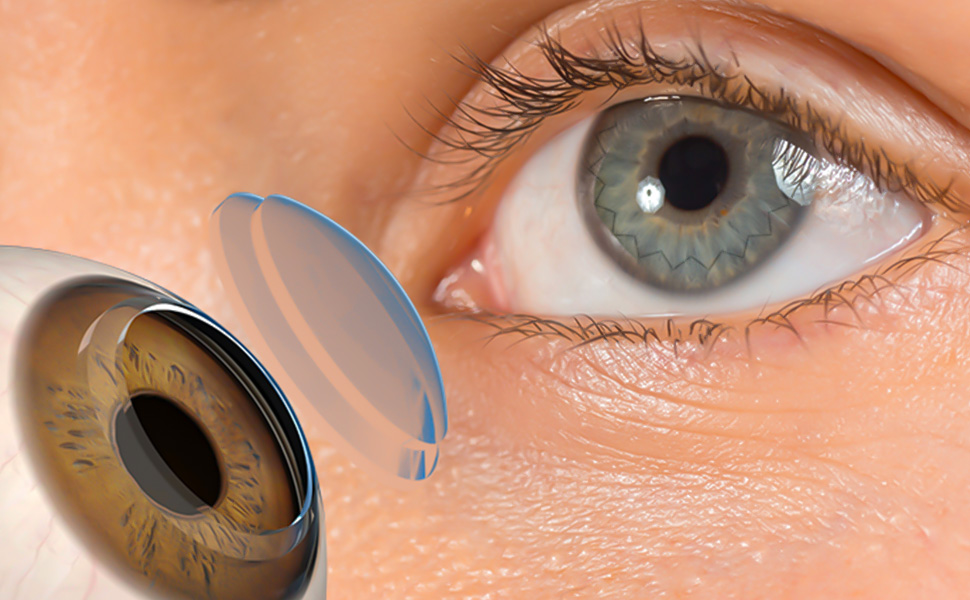
🔹 Keratoplasty (Corneal Transplant)
The most effective method for microcornea treatment is keratoplasty. In this surgical procedure, the damaged corneal layer is removed and replaced with healthy donor tissue.
Two types of keratoplasty can be performed:
-Full-thickness keratoplasty
-Lamellar (partial) keratoplasty
🔹 Surgery Process
-It is usually performed under local anesthesia (in some cases, general anesthesia is preferred).
-It takes 1–2 hours on average.
-The transplanted tissue is fixed with thin stitches.
-Vision recovery after surgery may vary from a few weeks to a few months.
Things to Consider After Surgery
-The eye may remain covered with a bandage for a few days.
-Your doctor will prescribe antibiotic and steroid drops to prevent infection.
-The eyes should not be rubbed and should be protected from foreign objects.
-Going to regular check-ups is very important for the healing process.
-Your doctor will prescribe antibiotic and steroid drops to prevent infection.
-The eyes should not be rubbed and should be protected from foreign objects.
-Going to regular check-ups is very important for the healing process.
Microcornea Prognosis (Course of Disease)?
With treatment, significant improvement in vision can be achieved in most patients.
-The success rate is between 70–90% in full-thickness and lamellar keratoplasty procedures.
-In mild cases, results that improve the quality of life can be obtained.
-In more severe cases, it is possible to reduce sensitivity to light and improve vision to some extent.
-The success rate is between 70–90% in full-thickness and lamellar keratoplasty procedures.
-In mild cases, results that improve the quality of life can be obtained.
-In more severe cases, it is possible to reduce sensitivity to light and improve vision to some extent.